Microtubules the seat of Consciousness

I found this statement on a science forum in regard to the chemical mechanism that responds to stimulus by producing neural “action potentials” that trigger response behaviors.

“They can only follow a very complex, yet technically predictable formula through sets of chemical reactions.”
What are human emotions?

Perhaps human emotions are triggered by a feed-back loop that in self-aware humans becomes an conscious emotional experience.

There is a clinical condition named Pseudobulbar Affect (BPA) that occurs when this feedback loop is constant instead of a single event.

What is pseudobulbar affect (PBA)?

Pseudobulbar affect (PBA) is a neurological condition that causes outbursts of uncontrolled or inappropriate laughing or crying. It is also known by other names including emotional lability, pathological laughing and crying, involuntary emotional expression disorder, compulsive laughing or weeping, or emotional incontinence. PBA is sometimes incorrectly diagnosed as a mood disorder – especially depression or bipolar disorder.

more…

What causes pseudobulbar affect (PBA)?

It is not completely known why pseudobulbar affect (PBA) occurs, but it is essentially always associated with neurological disorders or diseases that cause brain damage or injury. Disorders, diseases, or injuries that are associated with PBA include:

PBA occurs when there is a lack or loss of voluntary control over emotional responses. Various brain regions along a cerebro-ponto-cerebellar pathway are likely responsible for a loss of inhibitory or regulatory control on expression of emotions. Part of this pathway includes the cerebellum, which plays a key role in modulating or monitoring emotional responses and ensures they are appropriate to the social situation. Disruption of the neural (nerve) pathways from certain areas of the brain to the cerebellum may lead to a loss or lack of control over emotional expression.

Neurotransmitters, such as serotonin, norepinephrine, dopamine, and glutamate, are also thought to play a role in PBA.

(Pseudobulbar Affect (PBA): Causes, Symptoms & Treatment)

Note the glaring omission of the actual transport and response mechanism that is causal to this condition and that this condition may be caused by “Microtubule catastrophe”.

You agree that plants have sentience? Dictionary meaning will suffice.
the quality of being able to experience feelings:

Unfortunately I don’t have the time to sit down with this, so just a quick observation.

I noticed, you never referred to our hands. Nor feedbacks in general.

If I’m not mistake they are present in ALL cells. Is that correct?

That’s all.

Okay.

No argument.

This sounds like a gotcha, more than a serious question.

I think the point you should stop and think about is:

Get caught up on the state of knowledge,
then your questions will have a chance to start making sense.

Not a gotcha. You choose to enter the conversation therefore the question was to establish what side of the fence you sit on.

This may be of interest.

What is consciousness? It is awareness of self, others and our surroundings. What is sentience? It is the ability to perceive, experience and feel things.

The concept of sentience is a key part of animal rights. This is because it is necessary for animals to be sentient in order to experience pain and distress and the reason why reasonable people oppose animal cruelty. It is also part of the concept of the sanctity of life.

Scientific research shows that plants possess these same attributes. Plants are conscious, sentient beings.

In 1973, The Secret Life of Plants, by Peter Tompkins and Christopher Bird documented experiments that showed plant sentience. The book gave many examples of plant responses to human care, their ability to communicate, their reactions to music, their lie-detection abilities and their ability to recognize and to predict. It was a best seller. However, despite quoting from a range of research, it was met with skepticism by many scientists and academics.

Since then, thousands of published studies present a body of evidence that plants are conscious, sentient beings.

KEEP READING ON ECO FARMING DAILY

PLANT SENTIENCE AND THE IMPOSSIBLE BURGER (ANDRÉ LEU)

Foremost amongst this new breed of scientists is Monica Gagliano, research associate professor in evolutionary ecology at the University of Western Australia. Gagliano has published numerous peer-reviewed scientific papers on how plants have a Pavlov-like response to stimuli. They can learn, remember and communicate to neighboring plants. She has pioneered the research field of plant bioacoustics. She has demonstrated that plants emit their own “voices” and, moreover, detect and respond to the sounds of their environments.

Gagliano and other researchers have shown that learning, communicating, feeling, smelling, hearing and decision-making are not the exclusive province of animals. Below is a brief summary of some of this research.

I remember a study on how trees defend against caterpillar infestation.

The experiment involved two trees, not in direct contact with each other.

One tree was infected with a batch of caterpillars. The other tree was left undisturbed.

A typical defense mechanism of a tree in combatting caterpillars is the production of tannin in its leaves.
While caterpillars can adjust their digestive chemistry to ingest tanning this process robs the caterpillar of a lot of energy and affects its growth, making it weaker and more vulnerable to predation.

One of the remarkable evolutionary adaptions of trees is that when they produce tanning they do so selectively forcing a caterpillar to constantly adjust its digestive chemistry as it travels from leaf to leaf and that stunts its growth and limits the amount of damage it can inflict on the tree.

OK , a remarkable defensive strategy to a direct physical threat. But there’s more!

To the astonishment of the researchers, the other uninfected tree also began to produce tannin in a preemptive defensive strategy.

How did it know the proximity of a threat ?

The experiment was repeated several times and each time the uninfected tree began to produce tannin in response to the infected tree’s defensive behavior.

It was decided that trees can communicate via chemical signaling, much like bacteria communicate via quorum sensing.

Being that all trees contain microtubules in their leaf cells, it is entirely reasonable to assign some form of communication among leaves of the same tree as well as with leaves of adjacent trees.

Yes, microtubules, in addition to being the “information highways” in cells are the fundamental structural organelles in cytoplasma and cytoskeleton of all Eukaryotic organisms.

And it is not like each cell has a few microtubules, each cell is actually held together yet offer plasticity by hundreds , if not thousands of microtubules.

Microtubules in Plants

Abstract

Microtubules (MTs) are highly conserved polar polymers that are key elements of the eukaryotic cytoskeleton and are essential for various cell functions. αβ-tubulin, a heterodimer containing one structural GTP and one hydrolysable and exchangeable GTP, is the building block of MTs and is formed by the sequential action of several molecular chaperones. GTP hydrolysis in the MT lattice is mechanistically coupled with MT growth, thus giving MTs a metastable and dynamic nature. MTs adopt several distinct higher-order organizations that function in cell division and cell morphogenesis. Small molecular weight compounds that bind tubulin are used as herbicides and as research tools to investigate MT functions in plant cells. The de novo formation of MTs in cells requires conserved γ-tubulin-containing complexes and targeting/activating regulatory proteins that contribute to the geometry of MT arrays.

Various MT regulators and tubulin modifications control the dynamics and organization of MTs throughout the cell cycle and in response to developmental and environmental cues. Signaling pathways that converge on the regulation of versatile MT functions are being characterized.

INTRODUCTION

As early as the beginning of the 20th century, fibrous, filamentous, or tubular structures of similar diameter were described in various dividing eukaryotic cells. In the 1960s, high-resolution transmission electron microscopy images of these structures were obtained and it was determined with confidence that these filaments were actually hollow tubes, thus giving rise to the unified term “microtubules (MTs)” (Slautterback, 1963; Ledbetter and Porter, 1963).

and

Microtubule networks for plant cell division

Abstract

During cytokinesis the cytoplasm of a cell is divided to form two daughter cells. In animal cells, the existing plasma membrane is first constricted and then abscised to generate two individual plasma membranes. Plant cells on the other hand divide by forming an interior dividing wall, the so-called cell plate, which is constructed by localized deposition of membrane and cell wall material. Construction starts in the centre of the cell at the locus of the mitotic spindle and continues radially towards the existing plasma membrane.

Finally the membrane of the cell plate and plasma membrane fuse to form two individual plasma membranes. Two microtubule-based cytoskeletal networks, the phragmoplast and the pre-prophase band (PPB), jointly control cytokinesis in plants. The bipolar microtubule array of the phragmoplast regulates cell plate deposition towards a cortical position that is templated by the ring-shaped microtubule array of the PPB.

In contrast to most animal cells, plants do not use centrosomes as foci of microtubule growth initiation. Instead, plant microtubule networks are striking examples of self-organizing systems that emerge from physically constrained interactions of dispersed microtubules. Here we will discuss how microtubule-based activities including growth, shrinkage, severing, sliding, nucleation and bundling interrelate to jointly generate the required ordered structures.

Evidence mounts that adapter proteins sense the local geometry of microtubules to locally modulate the activity of proteins involved in microtubule growth regulation and severing. Many of the proteins and mechanisms involved have roles in other microtubule assemblies as well, bestowing broader relevance to insights gained from plants.

There are more microtubules on earth than all cells of all Eukaryotic organisms by a factor of perhaps 1000 . Perhaps only the water molecule H2O is more abundant than MT in biological cells.


Cell Nucleus in blue, Microtubule filaments in red.

https://v15.proteinatlas.org/learn/dictionary/cell/cytoskeleton+(microtubule+end)+1

I’m not on any side of the fence, beyond respecting physical reality and evolution.
“Consciousness” is a word that defines an infinite range of awareness, just as “love” is a word that defines an infinite range of affection, so is ultimately meaningless when it’s just tossed out as a parry.

Check out the article that Write shared:

Then ask a more nuanced question.


At least!
How could it be otherwise, they are a fundamental component of all eukaryotic cells?

Perhaps beyond their structural importance, there is also an element of being aware, or mirroring,
and as such they would be a fundamental component (building block?) of awareness, that with increasing complexity leads to consciousness and self-reflection. Perhaps microtubules will turn out to contain more variation and specialization than we are aware of presently, with a structural category and a memory/recall category, or something like that.

In any event, they are microscopic components, and will need various levels of orchestration and management, as we move up the evolutionary sequence of complexity - cellular awareness, to simple organisms, to creatures and then ever more complex on “up” to mammals with their incredible variety of awareness modes with us humans taking the most improbable twist of fate.

Beyond that, the only way to understand that increasing complexity of consciousness is to do a better job of incorporating the lessons of Evolution. …

1 Like

Hey Cc check out this paper where it concludes

This paper presents new arguments against plant consciousness, the most important of which are:

  1. A.Plants do not show proactive behavior.
  2. B.Classical learning does not indicate consciousness, so reports of such learning in plants are irrelevant.
  3. C.The considerable differences between the electrical signals in plants and the animal nervous system speak against a functional equivalence. Unlike in animals, the action potentials of plants have many physiological roles that involve Ca2+ signaling and osmotic control; and plants’ variable potentials have properties that preclude any conscious perception of wounding as pain.
  4. D.In plants, no evidence exists of reciprocal (recurrent) electrical signaling for integrating information, which is a prerequisite for consciousness.
  5. E.Most proponents of plant consciousness also say that all cells are conscious, a speculative theory plagued with counterevidence.

In furtherance of establishing serious interest by respected physicists, this is what intrigued Roger Penrose (Nobel laureate) when Stuart Hameroff (practising anesthesiologist) told him that it is the microtubules that respond to anesthetics by rendering the patient “unconscious”, but not affecting their function in maintaining homeostasis.

IOW, anesthetics don’t kill you, they just render you unconscious and unable to feel pain, etc.
But other than that they do not interfere with keeping all other homeostatic controls functional.
Moreover, it takes the exact same mixture of anesthetics to render all organisms unconscious and proves that microtubules perform the same functions in all conscious animals.

No one seems to be able to figure out how consciousness happens. That’s why Penrose suggests that needed to look deeper, but he did not have a vehicle that works at quantum scales until Hameroff suggested that microtubules might be the vehicle he was looking for.

Roger Penrose On Why Consciousness Does Not Compute - (in a conventional sense)
Nautilus | Science Connected

Penrose’s theory promises a deeper level of explanation. He starts with the premise that consciousness is not computational, and it’s beyond anything that neuroscience, biology, or physics can now explain. “We need a major revolution in our understanding of the physical world in order to accommodate consciousness,” Penrose told me in a recent interview. “The most likely place, if we’re not going to go outside physics altogether, is in this big unknown—namely, making sense of quantum mechanics.”

He draws on the basic properties of quantum computing, in which bits (qubits) of information can be in multiple states—for instance, in the “on” or “off” position—at the same time. These quantum states exist simultaneously—the “superposition”—before coalescing into a single, almost instantaneous, calculation. Quantum coherence occurs when a huge number of things—say, a whole system of electrons—act together in one quantum state.

It was Hameroff’s idea that quantum coherence happens in microtubules, protein structures inside the brain’s neurons. And what are microtubules, you ask? They are tubular structures inside eukaryotic cells (part of the cytoskeleton) that play a role in determining the cell’s shape, as well as its movements, which includes cell division—separation of chromosomes during mitosis. Hameroff suggests that microtubules are the quantum device that Penrose had been looking for in his theory. In neurons, microtubules help control the strength of synaptic connections, and their tube-like shape might protect them from the surrounding noise of the larger neuron. The microtubules’ symmetry and lattice structure are of particular interest to Penrose. He believes “this reeks of something quantum mechanical.”

How is it possible that single cells can communicate without a neural network?
There are many neuronless single-celled organisms that perform extraordinary feats of “logical” response behaviors to external stimulation. Cells have memory!

The Slime Mold is one such creature.

continued…

Here is the description of an organism that is little more than a collection of microtubules

The Slime mold, a single-celled organism that has no brain and no neural network, but it has a cytoskeleton and is filled with cytoplasm both organized by microtubules.

On the role of the plasmodial cytoskeleton in facilitating intelligent behavior in slime mold Physarum polycephalum

Abstract

The plasmodium of slime mold Physarum polycephalum behaves as an amorphous reaction-diffusion computing substrate and is capable of apparently ‘intelligent’ behavior. But how does intelligence emerge in an acellular organism?

Through a range of laboratory experiments, we visualize the plasmodial cytoskeleton—a ubiquitous cellular protein scaffold whose functions are manifold and essential to life—and discuss its putative role as a network for transducing, transmitting and structuring data streams within the plasmodium. Through a range of computer modeling techniques, we demonstrate how emergent behavior, and hence computational intelligence, may occur in cytoskeletal communications networks. Specifically, we model the topology of both the actin and tubulin cytoskeletal networks and discuss how computation may occur therein.

Furthermore, we present bespoke cellular automata and particle swarm models for the computational process within the cytoskeleton and observe the incidence of emergent patterns in both. Our work grants unique insight into the origins of natural intelligence; the results presented here are therefore readily transferable to the fields of natural computation, cell biology and biomedical science. We conclude by discussing how our results may alter our biological, computational and philosophical understanding of intelligence and consciousness.

Introduction

Slime mold Physarum polycephalum’s vegetative life cycle stage, the plasmodium (plural plasmodia, Fig. 1), is a macroscopic, multinucleate, acellular organism which behaves as a living amorphous reaction-diffusion computing substrate.[1]

To delineate, slime mold may be considered as an unconventional computing substrate in which data are represented as transitions in chemical equilibria in an excitable medium. The plasmodium is able to concurrently sense input from a range of stimuli including temperature, light, chemicals, moisture, pH and mechanical force.2-5 P. polycephalum’s innate behavior patterns may be manipulated experimentally to perform useful computational tasks, such as to calculating the shortest route between any number of spatially distributed nutrient sources, or navigating its way out of a maze via chemotaxis; these operations may be interpreted in terms of computational geometry, logic and spatial memory.[6,7]

image
Figure 1.
Plasmodium of slime mold P. polycephalum growing on an agar-filled Petri dish, feeding on porridge oats. Note the differences in morphology between medial/posterior plasmodial ‘veins’ (black arrow) and the ‘fan-shaped’ anterior margin (white arrow).

We are rapidly approaching the physical limitations of the materials used in the creation of traditional solid state computers, and their manufacture is ecologically-damaging. For these reasons, research into unconventional computing is gathering momentum. Unconventional, or non-classical computation, (UC) utilises the natural properties and processes of physical or living materials to provide useful computational functions. These systems are typically composed of simple and plentiful components, contain redundant parts (i.e. not being dependent on highly complex units), and show resilient or ‘fault tolerant’ behavior. UC is often observed in systems which show ‘emergent behavior’, novel behavior which emerges from the interactions between simple component parts and which—critically—cannot be described in terms of the lower level component interactions. Emergent behavior is found in systems with many simple, local interactions and which display self-organization, i.e. the spontaneous appearance of complexity or order from low-level interactions. Many of the attractive features of UC computing devices (distributed control, redundancy, fault tolerance) are generated by mechanisms of self-organization and emergence, and the study of these properties is useful not only from a computational perspective, but also from a biological viewpoint—since much of the complexity in living systems appears to be built upon these principles.

We begin by proposing that the P. polycephalum plasmodium must possess some form of network through which sensory and motor data may travel, as information transmission is a fundamental component of every system capable of computation (partnered with the means for data storage and logical processing of data). Furthermore, without such a network, incoming data—defined here loosely as an environmental stimulus which elicits a response within the organism—would be ‘unstructured’, data structuring here meaning the production of predictable, quantifiable data patterns from unstructured environmental data.

Indeed, in Ref.15, Lungarella and Sporns argue that the dynamical coupling of sensorimotor data streams with the morphology (here meaning both structure and physical properties) of a computational entity’s (biological or artificial) data network induces automatic information structuring, which in turn allows the entity to dynamically react to its environment: this is thought to be the underlying basis of learning and logical ability.[16-19]

Consequently, as the morphology of these data streams define how the entity may sense and interact with its environment, it essentially carries out a proportion of the computation. As such, computational processes are ‘outsourced’ to the morphology automatically, which reduces the workload of the entity’s control unit/s.[15,16,20-22]

This view of natural computing is derived from the precepts of ‘morphological computation’, a concept usually employed in robot design, wherein entity compliance is exploited to create self-stabilizing systems which reduce the need for constant monitoring by the control unit.[16,22]

By amalgamating biological and computer sciences, recent advances in the field have shown promise in modeling natural systems which have not been fully described in purely mathematical (algorithmic) terms, e.g. brain function.[23]

While assigning a tangible structure to the abstract term ‘data stream’ may appear to be a simplification for the ease of description, it is clear that some form of network for optimizing intracellular communication is present within slime mold, as the phenomena we may label as ‘inputs’ (i.e. environmental sensing) display redundancy, rapidity and transduction into different formats. For example, cellular signaling events may be observed to propagate far faster than simple diffusion of signal molecules through the cytoplasm would allow for, and many stimuli are transduced into multiple different formats, e.g., mechanical stimulation of a cell can provoke the generation of biochemical and bioelectrical signaling events.[24]

Slime molds would also appear to have a unique necessity for a data network as a single plasmodium can contain many millions of nuclei. While likening the functions of a cell’s nucleus with those of a processor is a contentious issue (which is not discussed here), it is clear that the nuclear processes which alter the cell/the cell’s behavior (activation/repression of genes, induction of signaling cascades etc.) are direct consequences of the environmental data it receives. How, then, are the activities of millions of nuclei are synchronized to produce coherent behavior? The existence of a plasmodial data network would appear to be an elegant solution to this problem, if such a network were demonstrated to connect nuclei together (hence facilitating internuclear communications) and/or interconnectivity (to allow for signal amplification).

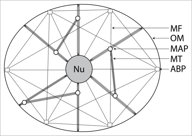
All eukaryotic (and some prokaryotic) organisms possess an intracellular network; a plentiful protein scaffold known as the cytoskeleton, which is composed of tubulin microtubules (MTs), actin microfilaments (MFs) and a range of intermediate filaments (IFs) (Fig. 2). Tubulin and actin are ubiquitous, while IF type differs depending on the function of the cell. All three cytoskeletal protein groups are considered to interlink with each other and with most of the major cellular organelles and receptors, to form one single interconnected network.[25]

The cytoskeleton participates a multitude of cellular functions, many of which are considered to be essential to life; these include mechanical rigidity, motility and substance (organelle and molecular) trafficking.[26]

image
Figure 2.
Simplified schematic representation of the cellular cytoskeleton in a generalized eukaryotic cell, illustrating how several varieties of cytoskeletal protein form an interconnected network which links cellular organelles together. Intermediate filaments and centrosome are not shown. Nu: nucleus; OM: outer membrane; MAP: microtubule-associated protein; MT: microtubule; ABP: actin-binding protein.

It is becoming increasingly evident that the cytoskeleton also participates in a wide range of cellular signaling events. The forms of information it may transmit include electrical potential, mechanical force, quantum events, propagating waves of protein conformational changes, facilitated chemical transport and biochemical signal transduction cascades.[24,26-32]

The cytoskeleton therefore represents an attractive model for describing data transmission within cells, as all of these signaling events may be interpreted as data being fed into a cellular computer. We are by no means the first to suggest this, but such a topic has not been previously described in slime mold models.

This study was undertaken to explore the putative role of the slime mold cytoskeleton in facilitating intelligent behavior by acting as an intraplasmodial data network. This was achieved by visualizing the plasmodial cytoskeleton with confocal microscopy and basing a range of computer modeling techniques on the structural observations made.

Results

Visualizing and formalising the plasmodial cytoskeleton

The actin and tubulin components of the P. polycephalum cytoskeleton were visualized with confocal microscopy, see Figures 3 and 4 (see Methodology section for details of sample preparation). Samples were taken from the 2 distinct anatomical locations of the plasmodium, namely the tubular plasmodial ‘veins’ that form the posterior areas of the plasmodium and the ‘fan-shaped’ advancing anterior margin formed from converging pseudopodia (see Fig. 1). The actin and tubulin components of the plasmodial cytoskeleton appear to be extremely complex, highly interconnected networks – especially so when compared to animal cell counterparts. The actin MF network is extremely abundant in the advancing pseudopodia (likely to result from the propulsion of motile machinery to the pseudopodial growth cone to facilitate movement), although somewhat less so in plasmodial veins (data not shown); the tubulin network would appear to be profuse in both anatomical locations, but less so than actin in the anterior portions of the organism. Both cytoskeletal proteins appear to articulate onto each nucleus at several points and are also present at the external membrane, indicating associations with surface receptors (as has been demonstrated in animal cells).26

more…

What does this suggest about microtubules???

Moreover, I can see no more persuasive argument that microtubules are the prime constituent of “intelligent networks” than that even single-celled brainless and neuronless organisms have inherent “communication” skills that may be very much like “quorum sensing” in bacteria, plants and eventually acquire emergent self-aware conscious intelligence in more evolved complex animals.

An organism does not need to be conscious to be sentient and responsive to survival demands.

Consciousness is the evolutionary finish of increasingly complex sensory response networks and emerges at every level of natural selection of survival strategies.

From you paper:

Finally, we present our own hypothesis, based on two logical assumptions, concerning which organisms possess consciousness.

Our first assumption is that affective (emotional) consciousness is marked by an advanced capacity for operant learning about rewards and punishments.

Our second assumption is that image-based conscious experience is marked by demonstrably mapped representations of the external environment within the body.

Certain animals fit both of these criteria, but plants fit neither. We conclude that claims for plant consciousness are highly speculative and lack sound scientific support.

Now the question is, how reasonable are their assumptions? I don’t pretend to know the answer, just saying if they don’t delve into the evolution of the full spectrum of consciousness, they are missing most the show.

You might to google “Are single celled creatures conscious?” for a selection of some provocative reading.

It has also been identified that three cellular structures and mechanisms that likely play critical roles here are excitable membranes, oscillating cytoskeletal polymers, and structurally flexible proteins. Finally, basic biophysical principles are proposed to guide those processes that underly the emergence of supracellular sentience from cellular sentience in multicellular organisms.

{ What do you think Write, are those your microtubules? }

This one’s fun:


Re 150, Write,
It is fascinating, no doubt.
Next few years should be amazing.

Getting a better understanding of how those tiniest of components build up in size and complexity.

1 Like

Oh, yes.

To follow the gradual emergent refinement of sensory abilities and response behaviors along with the increasing complexity in microtubular network growth in all Eukaryotic biology based on cytoplasmic and cytoskeletal cells.

It does not need to be conscious to be logically intelligent, but it must be logically intelligent to also acquire consciousness.

Classic. Its your paper

citizenschallengev4

1d

This sounds like a gotcha, more than a serious question.

I think the point you should stop and think about is:

That’s a quote from the paper. That’s what we do here, we speculate. As long as you say you are speculating, and present data to show why you are speculating, and present your own thoughts, then this can be a fun place to explore ideas.

Which means we don’t need to agree, but we should both display some good faith.

Such as:
(1) plants have not been shown to perform the proactive, anticipatory behaviors associated with consciousness, but only to sense and follow stimulus trails reactively;
(2) electrophysiological signaling in plants serves immediate physiological functions rather than integrative-information processing as in nervous systems of animals, giving no indication of plant consciousness;
(3) the controversial claim of classical Pavlovian learning in plants, even if correct, is irrelevant because this type of learning does not require consciousness.

Our first assumption is that affective (emotional) consciousness is marked by an advanced capacity for operant learning about rewards and punishments.
Our second assumption is that image-based conscious experience is marked by demonstrably mapped representations of the external environment within the body.

Certain animals fit both of these criteria, but plants fit neither.

We conclude that claims for plant consciousness are highly speculative and lack sound scientific support.

What I see is that they set very specific parameters and conclude anything outside of that has zero consciousness. I think that’s jumping to conclusions.
I’m thinking they are perhaps missing a few things worth thinking about, with their narrow constraints.

This is how I see it from an evolutionary perspective.

Evolution of Microbial Quorum Sensing to Human Global Quorum Sensing: An Insight into How Gap Junctional Intercellular Communication Might Be Linked to the Global Metabolic Disease Crisis

by
James E. Trosko
Department of Pediatrics/Human Development, College of Human Medicine, Michigan State University, East Lansing, MI 48824, USA

…, since the mechanism of cell communication itself is universal in biology, in keeping with a Kuhnian paradigm shift. This approach may even elucidate the nature and evolution of consciousness as a manifestation of the cellular continuum from unicellular to multicellular life. We need such a functional genomic mechanism for the process of evolution if we are to make progress in biology and medicine.” John S. Torday, Prospect Biol. Med. 56: 455, 2014. "

While the start of this analysis must begin with very few facts about the first set of molecular events to create the first living, self-replicating cell, it is clear that many independent events must have occurred to create a small space in that vast primordial sea, in which the “inside” was separated from the “outside.” This implies the formation of a semi-permeable membrane. In addition, that “inside space” had to facilitate the movement of ions/small molecules, in and out of that space, while at the same time allowing for the creation of self-replicating molecules that code for all the components to allow this unique separated space to maintain “homeostatic control of all the necessary biochemical functions” to allow this cell to generate energy for life and reproduce its survival machinery in an ever-changing world.

It is not to be underestimated that our understanding of those early events is, today, still unknown. However, it must have happened because before that “moment” in history, there was no living organism. In addition, in keeping with this analysis, it occurred within natural physical and chemical scientific processes, not by “divine intervention” or by “vital forces.”

From the beginning of that moment to this day, when there are seven billion human beings on earth surrounded by untold numbers of other micro- and macro-species, an environment is being depleted of vital factors needed for basic survival, as well as polluted with the degradable and undegradable detritus of the unconscious and conscious, culture-building Homo sapiens.

It is necessary to keep in mind the continuous and intimate interconnectedness of life to ponder how the current situation on Earth, including the continuous loss of species and their state of health, might be associated with the evolution of communication: first “quorum-sensing” [3] between micro-organisms, then chemical communication between unconscious plants, and on to various forms of primitive “languages” of multi-cellular metazoans (visual-based body language and body phenotypes; tactile, sound or voice communication, etc.) and consciousness of one’s individual consciousness via gap junctional intercellular communication (GJIC).

While it might seem outrageous to single out the family of connexin or gap junction genes from the approximately 25 thousand genes in the human genome that led to (a) symbols of conscious objects; (b) translation of those abstractions so a spoken language could be communicated; (c) the transformation of those abstract symbols or ideas into technologies; and (d) finally, the ability to decide or “value” whether to apply that technology for life and reproduction [4], the premise is based on the fact that, without those early acquired genes, none of the differentiated cells, such as neurons and the functional brain, would exist.

This concept of Homo sapiens’ state of evolution lets us see that “culture” has emerged as an evolutionary process via cybernetic and hierarchical principles [5,6].

An organism can be sentient, i.e. have sensory responses, without having consciousness.

This process is also present in human homeostasis, where the body (and brain) act as an unconscious control system rather than making conscious decisions about what hormones to release in response to internal stimulus. (Interoception, Anil Seth)

IMO, the mechanism that allows for unconscious quorum sensing is the microtubular network in the cell’s cytoskeleton and cytoplasm and acts as an EM hive mind responsible for intra-cellular and inter-cellular information distribution within and between cells.

As this self-organizing information micro-processor is a common denominator in all Eukaryotic organisms it should be the best candidate as the substrate for the emergence of self-aware consciousness in brained organisms.

Continuing in our discovery of microtubular functions.

The Emerging Complexity of the Vertebrate Cilium: New Functional Roles for an Ancient Organelle

Cilia and flagella are found on the surface of a strikingly diverse range of cell types. These intriguing organelles, with their unique and highly adapted protein transport machinery, have been studied extensively in the context of cellular locomotion, sexual reproduction, or fluid propulsion. However, recent studies are beginning to show that in vertebrates particularly, cilia have been recruited to perform additional developmental and homeostatic roles.

Here, we review advances in deciphering the functional components of cilia, and we explore emerging trends that implicate ciliary proteins in signal transduction and morphogenetic pathways.

Ciliary structure is strongly conserved from protists to multicellular organisms. The existence of 9+2 organelles across eukaryotes has led to the speculation that this may be the ancestral structure, with 9+0 cilia having arisen later (Cavalier-Smith, 2002, Mitchell, 2004). A traditional view has maintained that cilia with the central pair of microtubules, such as sperm flagella, oviduct, and respiratory tract cilia, are motile, whereas their absence is indicative of nonmotile sensory cilia, as exemplified by renal, pancreatic, photoreceptor, and olfactory neuron cilia in vertebrates.

However, several notable exceptions, such as the 9+0 microtubule structure in diatom gamete flagella (Manton et al., 1970) and eel sperm flagella (Gibbons et al., 1985), suggest that this additional pair of microtubules is not a prerequisite for motility. In the mouse, 9+0 cilia generate fluid flow in the embryonic node (Nonaka et al., 2002), while, in zebrafish, motile 9+0 cilia have been observed in the central canal of the spinal cord (Kramer-Zucker et al., 2005).

Cilia and Motility

Motility is the function most commonly associated with cilia, and in 9+2 cilia, the central microtubule pair appears to be necessary to confer movement via its dynein-mediated link with the surrounding outer doublets (Satir, 1999). The mechanical constraints given by the linkage between dynein arms, microtubule doublets, and radial spokes then convert interdoublet sliding into characteristic ciliary movement. The precise pattern of movement of individual cilia has been studied extensively both on ciliated epithelia and in the flagella of the green alga Chlamydomonas. The beat is asymmetric, and it consists of an effective stroke occupying one-quarter of the beat and a recovery stroke. In wild-type flagella, the beat frequency is ∼60 Hz, and in flagella lacking outer arm dyneins, the beat form remains nearly normal; however, the frequency decreases to 20–30 Hz (Porter and Sale, 2000).

Coordinated beating between adjacent cilia, with each individual cilium having an inherently asymmetric beat, results in directional movement of extracellular fluid overlying ciliated epithelia such as those found in the trachea, choroid plexus, and oviduct. Recently, motile cilia in the ependyma of the brain were shown to be essential for directing long-range directional migration of neuroblasts (Sawamoto et al., 2006).

In addition to the classic functions of ciliary motility, vertebrates are thought to utilize the asymmetry inherent in the structure of the cilium, as well as the resulting asymmetric ciliary movement, as a means to generate handed left-right (LR) asymmetry during development (Tabin and Vogan, 2003). This process depends on a subpopulation of motile cilia not found on ciliated epithelia, specifically the monocilia located on the ventral cells of the mammalian node (organizer) and the epithelium lining Kupffer’s vesicle in fish (Amack and Yost, 2004, Essner et al., 2005, Kramer-Zucker et al., 2005, Nonaka et al., 1998).

Curiously, several reports show node monocilia that lack the central pair and radial spoke apparatus (Bellomo et al., 1996, Nonaka et al., 1998, Sulik et al., 1994). In addition, the beat pattern of node monocilia may deviate from that observed in Chlamydomonas and ciliated epithelia: node cilia have an effective recovery stroke like other motile cilia, but the motion is conical and the directionality of the resulting fluid movement may be, in part, secondary to a posterior tilt of the cilium itself relative to the cell body combined with the inherent asymmetry of the ciliary beat (Nonaka et al., 2005, Okada et al., 2005).

Furthermore, these cilia beat in a clockwise direction at frequencies slightly lower than those of ciliated epithelia (Nonaka et al., 1998, Okada et al., 2005, Sulik et al., 1994, Supp et al., 1999). Coordinated beating of >200 (in mouse) monocilia generates leftward directional movement of the extracellular fluid surrounding the node, called nodal flow (Nonaka et al., 1998, Okada et al., 1999).

Directional fluid flow is essential for the development of LR asymmetry; mice with paralyzed node monocilia due to a mutation in the axonemal dyneins lrd (Okada et al., 1999, Supp et al., 1999) and Dnahc5 (Olbrich et al., 2002) and zebrafish with morpholinos targeted against the zebrafish lrd ortholog (Essner et al., 2005) have absent nodal flow and randomization of LR asymmetry. The LR defect in lrd−/− mouse embryos is rescued by artificial leftward nodal flow, demonstrating that Lrd functions in LR development via its role in node-cilia motility (Nonaka et al., 2002).

The mechanism by which adjacent cells in the node or Kupffer’s vesicle coordinate ciliary movement to generate laminar directional fluid flow is poorly understood. One clue comes from mice with mutations in the ankyrin repeat-containing protein inversin (Inv): these mice have normal movement of the individual node cilium; however, nodal flow is sluggish, due, at least in part, to defective ciliary tilt, and LR development is abnormal (Okada et al., 1999, Okada et al., 2005). These data suggest that Inv may be required to coordinate movement of adjacent cilia.

Cilia as Chemosensory and Mechanosensory Organelles
https://www.sciencedirect.com/science/article/pii/S1534580706002772

In addition to the “known” role microtubules and associated filaments play in the brain, to me the above research confirms the important role microtubules play in the emergent phenomenon of consciousness.

There is no known systemic mechanism that offers so many dynamical action potentials as the microtubule networks in cellular cytology.

The fact that in addition to intra-cellular commuication and transportation, the inter-cellular communication and the ultimate collective processing of all sensory information by the brain, cries out for recognition of the sufficient dynamical processing that evokes an emergent experiential ability.

I imagine you’re kind of a, ‘I just read the articles’ kinda guy. Me what can I say, I like the pictures too.
:wink:
Though, indeed those articles are fascinating, until they make my head spin, then I gotta back off a little.

Figure 1. Gap junctions in cellular homeostasis. Extracellular signals, such as growth factors, cytokines, hormones, toxicants, extra-cellular matrices, and cell adhesion molecules, that vary for each cell type (adult stem cell, progenitor, and terminally differentiated), interact with receptor-dependent or receptor-independent targets, which then activate intracellular signal transduction pathways that induce the transcription of genes through activated transcription factors. These specific intracellular pathways operate under cascading systems that cross-communicate with each other in controlling the expression of genes that direct the proliferation, differentiation, and apoptosis of cells within a tissue. These multiple intracellular signaling check points are further modulated by intercellular signals traversing gap junctions, thereby maintaining the homeostatic state of a tissue. Abnormal interruption of these integrated signaling pathways by food-related and environmental toxins/toxicants will disrupt the normal homeostatic control of cell behavior (Permission granted from Toxicology2010, 270, 18–34 [22]).

https://ars.els-cdn.com/content/image/1-s2.0-S1534580706002772-gr1.jpg

Figure 1. Cilia Mediate Mechanosensation

Cilia are unique cellular structures that typically protrude from the apical surface of mammalian cells. Anchored to the basal body, which consists of nine radially oriented microtubule triplets, the cilium extends from the cell with nine microtubule doublets either without (9+0; shown) or with (9+2; not shown) a central pair of microtubules. Ciliary protein trafficking along the axoneme is facilitated by IFT rafts such as Polaris/IFT88 and is carried out in the anterograde direction by kinesin-2 molecular motors, whereas retrograde shuttling of proteins is conducted by dynein. A mechanosensory role has been demonstrated for cilia; more specifically, extracellular flow causes the cilium to bend and results in an increase of intracellular Ca2+.

Evidence suggests that the flow sensation in kidney primary cilia is mediated by PC1 and PC2, transmembrane proteins that colocalize to the ciliary membrane and form a cation-permeable channel. Figure adapted from Figure 1 (Guay-Woodford, 2003) with permission.

This may be of interest.

A narrative of microtubule sensory function in plants

Migration via quantum mechanics

Experiments have shown that changes in the magnetic field at only one thousandth of the strength of the Earth’s magnetic field can effect the orientation of the migratory European robin. Studies also suggest that entanglement can survive in the robin’s retina for up to 100 microseconds, compared to the best so far achieved, in super-cooled laboratory conditions, of only 80 microseconds.

From the point of view of quantum consciousness, the European robin research, along with research on photosynthetic organisms, is interesting in appearing to refute the fifteen-year-old and often trumpeted study of Max Tegmark purporting to show that quantum features could not be functional in biological matter.
American Physical Society Sites :: PhysicsCentral

While microtubules are nor specifically mentioned in this article, the prior article on microtubule function in the eye explains the necessity of microtubule processing of electromagnetic energy, allowing for the evolution of navigational skills in migrating birds.